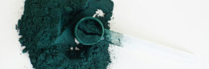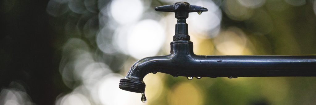
Cleaning water with silver
Silver nanoparticles have unique physical, chemical, and biological properties compared to their bulk parent materials, due to their high surface-to-volume ratio and the nanometer scale. Silver nanoparticles are toxic to microorganisms and the antibacterial effects1 (even against antibiotic-resistant strains2) and antiviral effects3 have been used to control pathogenic microorganisms to provide safe drinking water from contaminated sources4,5. Traditional disinfection methods, such as chlorine, can produce harmful by-products with high geno- and cytotoxicity6. However, it is important to also address the potential risks associated with exposure to silver nanoparticles in suspension, both to human health and the environment7. Immobilizing silver nanoparticles on supports, like cellulose8 or cotton fabric9, minimizes risks and enhances antibacterial activity. These support materials prevent agglomerate formation, enable nanoparticle recovery for reuse, and reduce synthesis and application costs10.
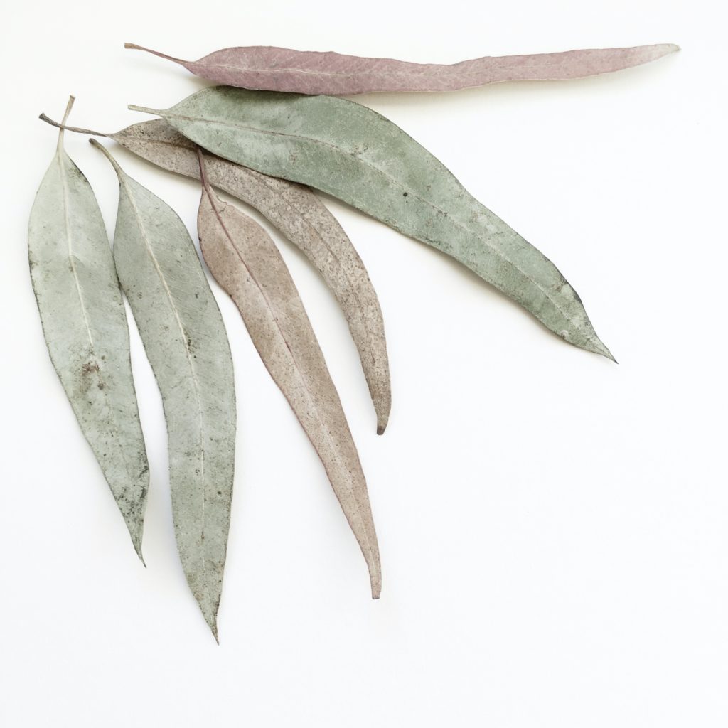
How to make silver cotton with Eucalyptus leaves
Nanoparticle synthesis can be achieved through different methods, utilizing chemical and physical pathways. These conventional methods are not considered environmentally friendly, as they consume high amounts of energy and use toxic and expensive solvents and reagents11. One approach to greener synthesis is the utilization of microorganisms or plant extracts12, which eliminates the requirement for high pressures, high temperatures, and toxic chemical reagents13. While the use of microorganisms is often challenging11, using plant biomass for nanoparticle synthesis provides an alternative that is effective and simple14. For silver nanoparticles, the first successful synthesis using leaf extracts was reported in 200315. Since then, researchers have explored a wide variety of plant extracts for this purpose16. Southern blue gum leaves, which are typically considered waste products in cellulose production17,18, contain phenolic compounds which makes them an excellent candidate for the production of silver nanoparticles.
Advancing synthesis by understanding the chemical processes
To improve green synthesis it is necessary to identify biomolecules or phytochemicals and understand their roles in the synthesis processes. Phytochemicals present in plant extracts play a crucial role in the synthesis of nanoparticles by acting as reducing and capping agents, ensuring their stability during formation19. Among these phytochemicals, phenolic compounds are primarily responsible for nanoparticle formation20, while carbohydrates, proteins, terpenoids, and vitamins also contribute to the process19,21. However, the specific roles of these phytochemicals in terms of nanoparticle stability, aggregation, morphology, and reactivity remain unclear16. By gaining a better understanding of these components, it becomes possible to enhance the efficiency and effectiveness of green synthesis methods.
Researchers from several Chilean universities have investigated the phytochemicals that are involved in the formation of silver nanoparticles on cellulose paper and cotton fabric supports using Eucalyptus extracts to develop an efficient and eco-friendly protocol22. The Eucalyptus leaves are washed, dried, crumbled, heated in water, centrifuged, and filtered to get a leaf extract. The paper and fabric is inserted into silver nitrate solution, then removed and washed and inserted into the plant extract to reduce silver ions. Also they test sodium hydroxide to modify the pH of the system and test the effect on the structure and properties of the resulting nanoparticle. Different from previous studies that focused only on qualitative spectrophotometric analyses, this research group used UHPLC-QTOF-MS to analyse the phytochemical content in the leaf extracts before and after the nanoparticles are produced.
Unveiling the role of biomolecules in silver nanoparticle synthesis
In the quest to understand the intricate process of silver nanoparticle synthesis, the identification of biomolecules involved in the transformation becomes paramount. Characterizing Eucalyptus leaf extracts before and after nanoparticle formation provides valuable insights. FTIR spectroscopy analysis indicated that phenolic compounds, reducing sugars, and proteins are involved in the synthesis process of silver nanoparticles. Spectrophotometric analysis further revealed that phenolic compounds decrease in concentration after silver nanoparticle formation, indicating their possible role as reducing agents or stabilizers on the surface of the nanoparticles. Concentration of reducing sugars decreased only slightly, implying a potential stabilizing role. The protein content did not exhibit considerable changes. The researchers used UHPLC-QTOF-MS to identify the key phenolic compounds involved in the silver reduction. Most of the phenolic compounds were reported in Eucalyptus leaves before23–25. The remaining features were annotated using SIRIUS CSI:FingerID26. The main phenolic compounds that act as reducing agents are those derived from gallic acid, mainly due to the large number of -OH groups in these compounds.
Overall they found that cotton fabric as support generates a more notable decrease in phenolic compounds in the leaf extracts and a higher proportion of nanoparticles on the fabric. Moreover, the silver nanoparticles on cotton fabric show excellent photocatalytic activity and antibacterial against E. coli and could be an interesting application for remediation of contaminated waters.
References
- 1.Tang S, Zheng J. Antibacterial Activity of Silver Nanoparticles: Structural Effects. Adv Healthcare Mater. Published online May 29, 2018:1701503. doi:10.1002/adhm.201701503
- 2.Agnihotri S, Sillu D, Sharma G, Arya RK. Photocatalytic and antibacterial potential of silver nanoparticles derived from pineapple waste: process optimization and modeling kinetics for dye removal. Appl Nanosci. Published online September 27, 2018:2077-2092. doi:10.1007/s13204-018-0883-9
- 3.El-Sheekh MM, Shabaan MT, Hassan L, Morsi HH. Antiviral activity of algae biosynthesized silver and gold nanoparticles against Herps Simplex (HSV-1) virus in vitro using cell-line culture technique. International Journal of Environmental Health Research. Published online July 6, 2020:616-627. doi:10.1080/09603123.2020.1789946
- 4.Kumar A, Mishra B, Tripathi BP. Polydopamine assisted synthesis of ultrafine silver nanoparticles for heterogeneous catalysis and water remediation. Nano-Structures & Nano-Objects. Published online July 2020:100489. doi:10.1016/j.nanoso.2020.100489
- 5.Furst KE, Pecson BM, Webber BD, Mitch WA. Tradeoffs between pathogen inactivation and disinfection byproduct formation during sequential chlorine and chloramine disinfection for wastewater reuse. Water Research. Published online October 2018:579-588. doi:10.1016/j.watres.2018.05.050
- 6.Zhao Y, Sun Y, Gao B, Wang Y, Yang Y. Inhibition of disinfection by-product formation in silver nanoparticle-humic acid water treatment. Separation and Purification Technology. Published online August 2017:158-167. doi:10.1016/j.seppur.2017.04.039
- 7.Agnihotri S, Mukherji S, Mukherji S. Immobilized silver nanoparticles enhance contact killing and show highest efficacy: elucidation of the mechanism of bactericidal action of silver. Nanoscale. Published online 2013:7328. doi:10.1039/c3nr00024a
- 8.Wu J, Zhao N, Zhang X, Xu J. Cellulose/silver nanoparticles composite microspheres: eco-friendly synthesis and catalytic application. Cellulose. Published online June 7, 2012:1239-1249. doi:10.1007/s10570-012-9731-3
- 9.Xing H, Cheng J, Tan X, Zhou C, Fang L, Lin J. Ag nanoparticles-coated cotton fabric for durable antibacterial activity: derived from phytic acid–Ag complex. The Journal of The Textile Institute. Published online September 26, 2019:855-861. doi:10.1080/00405000.2019.1668137
- 10.Agnihotri S, Mukherji S, Mukherji S. Impact of background water quality on disinfection performance and silver release of immobilized silver nanoparticles: Modeling disinfection kinetics, bactericidal mechanism and aggregation behavior. Chemical Engineering Journal. Published online September 2019:684-696. doi:10.1016/j.cej.2019.04.186
- 11.Huang J, Zhan G, Zheng B, et al. Biogenic Silver Nanoparticles by Cacumen Platycladi Extract: Synthesis, Formation Mechanism, and Antibacterial Activity. Ind Eng Chem Res. Published online July 1, 2011:9095-9106. doi:10.1021/ie200858y
- 12.Swilam N, Nematallah KA. Polyphenols profile of pomegranate leaves and their role in green synthesis of silver nanoparticles. Sci Rep. Published online September 9, 2020. doi:10.1038/s41598-020-71847-5
- 13.Mittal AK, Bhaumik J, Kumar S, Banerjee UC. Biosynthesis of silver nanoparticles: Elucidation of prospective mechanism and therapeutic potential. Journal of Colloid and Interface Science. Published online February 2014:39-47. doi:10.1016/j.jcis.2013.10.018
- 14.Mohanpuria P, Rana NK, Yadav SK. Biosynthesis of nanoparticles: technological concepts and future applications. J Nanopart Res. Published online August 3, 2007:507-517. doi:10.1007/s11051-007-9275-x
- 15.Shankar SS, Ahmad A, Sastry M. Geranium Leaf Assisted Biosynthesis of Silver Nanoparticles. Biotechnol Prog. Published online December 5, 2003:1627-1631. doi:10.1021/bp034070w
- 16.Vanlalveni C, Lallianrawna S, Biswas A, Selvaraj M, Changmai B, Rokhum SL. Green synthesis of silver nanoparticles using plant extracts and their antimicrobial activities: a review of recent literature. RSC Adv. Published online 2021:2804-2837. doi:10.1039/d0ra09941d
- 17.Andrade A, Henríquez-Gallegos S, Albornoz-Palma G, Pereira M. Effect of the chemical and structural characteristics of pulps of Eucalyptus and Pinus on the deconstruction of the cell wall during the production of cellulose nanofibrils. Cellulose. Published online April 17, 2021:5387-5399. doi:10.1007/s10570-021-03848-0
- 18.Andrade A, Herrera OE, Reyes-Contreras P, Pereira M, Vásquez-Garay F. Eucalyptus globulus bark valorization: Production of fibers by Neutral Sulphite Semi-Chemical Process for Liner Paper Manufacture. Floresta Ambient. Published online 2021. doi:10.1590/2179-8087-floram-2020-0048
- 19.Salgado P, Mártire DO, Vidal G. Eucalyptusextracts-mediated synthesis of metallic and metal oxide nanoparticles: current status and perspectives. Mater Res Express. Published online June 7, 2019:082006. doi:10.1088/2053-1591/ab254c
- 20.Vijayaraghavan K, Ashokkumar T. Plant-mediated biosynthesis of metallic nanoparticles: A review of literature, factors affecting synthesis, characterization techniques and applications. Journal of Environmental Chemical Engineering. Published online October 2017:4866-4883. doi:10.1016/j.jece.2017.09.026
- 21.El-Seedi HR, El-Shabasy RM, Khalifa SAM, et al. Metal nanoparticles fabricated by green chemistry using natural extracts: biosynthesis, mechanisms, and applications. RSC Adv. Published online 2019:24539-24559. doi:10.1039/c9ra02225b
- 22.Salgado P, Bustamante L, Carmona DJ, et al. Green synthesis of Ag/Ag2O nanoparticles on cellulose paper and cotton fabric using Eucalyptus globulus leaf extracts: Toward the clarification of formation mechanism. Surfaces and Interfaces. Published online August 2023:102928. doi:10.1016/j.surfin.2023.102928
- 23.Mandal D, Bolander ME, Mukhopadhyay D, Sarkar G, Mukherjee P. The use of microorganisms for the formation of metal nanoparticles and their application. Appl Microbiol Biotechnol. Published online November 30, 2005:485-492. doi:10.1007/s00253-005-0179-3
- 24.Boulekbache-Makhlouf L, Meudec E, Mazauric JP, Madani K, Cheynier V. Qualitative and Semi-quantitative Analysis of Phenolics in Eucalyptus globulus Leaves by High-performance Liquid Chromatography Coupled with Diode Array Detection and Electrospray Ionisation Mass Spectrometry. Phytochem Anal. Published online August 29, 2012:162-170. doi:10.1002/pca.2396
- 25.Boulekbache-Makhlouf L, Meudec E, Chibane M, et al. Analysis by High-Performance Liquid Chromatography Diode Array Detection Mass Spectrometry of Phenolic Compounds in Fruit of Eucalyptus globulus Cultivated in Algeria. J Agric Food Chem. Published online December 1, 2010:12615-12624. doi:10.1021/jf1029509
- 26.Dührkop K, Fleischauer M, Ludwig M, et al. SIRIUS 4: a rapid tool for turning tandem mass spectra into metabolite structure information. Nat Methods. Published online March 18, 2019:299-302. doi:10.1038/s41592-019-0344-8